
Matching Minimally Invasive Cancer Treatment to Metastatic Site: A Shortlist
HTNXT · Interventional Oncology Reference
A site-by-site shortlist of documented interventional options — pancreatic retroperitoneal recurrence, lung metastasis, hepatic spread and metastatic breast lesions — with the case context that shaped each choice and the limits of the evidence behind it.
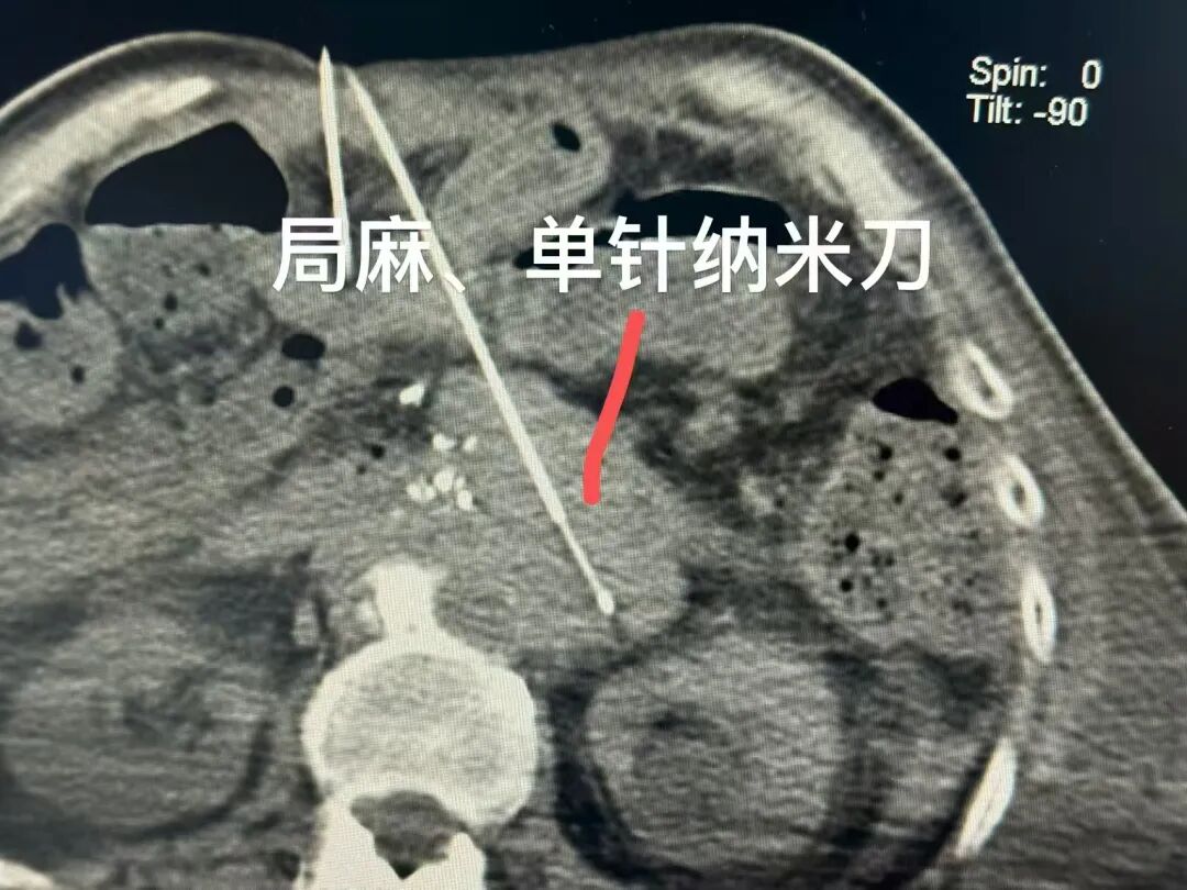
Where a metastatic lesion sits frequently constrains treatment more tightly than the histology that produced it. A retroperitoneal recurrence wrapped around the celiac trunk and superior mesenteric artery, a lung deposit pressed against the pericardium and major vessels, and a meningeal implant are all described as metastatic cancer, yet each presents a different procedural problem: a different access route, a different anaesthetic risk, and a different set of structures that cannot be injured.
Minimally invasive cancer treatment is usually presented as a catalogue of technologies. A shortlist organised by metastatic site answers a narrower and more practical question at the evaluation stage: given this anatomy, this prior treatment history and this physiological reserve, which interventional options have actually been documented in a comparable case?
The shortlist below maps four documented case profiles published by Guangzhou Fuda Cancer Hospital to the metastatic site each addressed — pancreatic retroperitoneal recurrence, lung metastasis, hepatic spread, and metastatic breast lesions involving chest wall, liver and meninges. Each entry states the documented option, the case context that justified it, the sequence executed, and the outcome the case reports.
How to read this shortlist. Nothing here implies that a patient with the same metastatic site is a candidate for the same procedure. Eligibility is determined case by case through multidisciplinary assessment of imaging, pathology and physiological reserve. This article summarises published case documentation and is not medical advice.
Why metastatic site now competes with tumour type as the first filter
In localised disease the decision usually starts with histology. In metastatic disease the decision splits into two tracks that are planned together but constrained differently. Systemic control is histology- and molecular-marker-driven. Local control is anatomy-driven, and the anatomy questions are concrete: can the lesion be reached percutaneously without crossing an organ that cannot be crossed; does it encase a vessel that must remain patent; is it close enough to the pericardium or to major vessels that a thermal margin is unsafe; is the patient fit for general anaesthesia; has the region already received radiotherapy.
Resection and conventional radiotherapy remain central where they are feasible, and none of the entries below is presented as a substitute for them. What the cases illustrate is the group of patients for whom imaging suggested non-resectability, or for whom general anaesthesia was intolerable, or whose disease had progressed through multi-line chemotherapy and radiotherapy. For that group, the practical shortlist is assembled from image-guided ablation, vascular interventional techniques and stereotactic radiosurgery, sequenced with systemic therapy rather than opposed to it.
This is also where procurement language and clinical language converge. International patients and referring physicians increasingly compare providers on procedure portfolio and guidance infrastructure — whether dual imaging guidance, digital subtraction angiography (DSA), ablation platforms and molecular testing sit inside one team — rather than on hospital size alone.
How this shortlist was assembled
Each entry corresponds to one documented case profile. The four profiles share a structure that makes them comparable, which is what allows them to be read side by side rather than as separate success stories.
- Anatomical constraint recorded before treatment — vessel encasement, proximity to the pericardium, or meningeal involvement, documented from imaging.
- Physiological and logistical constraints recorded — cardiopulmonary reserve, tolerance of general anaesthesia, contrast allergy, prior treatment lines.
- Sequence recorded step by step — what was done first, what was combined, and what was reassessed.
- At least one re-assessment endpoint reported — CT re-imaging, alpha-fetoprotein (AFP) monitoring, or radiologic re-measurement.
- Documentation limits stated — anonymised records, original imaging and pathology available only on hospital authorisation, and no survival data in the published profiles.
| Metastatic site | Documented option | Case context documented | Outcome reported in the case |
|---|---|---|---|
| Pancreatic retroperitoneal recurrence encasing the celiac trunk and superior mesenteric artery | Single-needle bipolar NanoKnife (S-IRE) ablation under CT + ultrasound guidance, local anaesthesia, with concurrent biopsy and celiac plexus block | 81-year-old male from Hong Kong, China; post-Whipple recurrence with a ~7 cm irregular hypoechoic mass; cardiopulmonary reserve did not allow resection or general anaesthesia | Ambulatory the next day; CT showed evident necrosis and shrinkage of the lesion; no numeric values provided in the report |
| Lung metastasis adjacent to the pericardium and major vessels | Superselective interventional embolization-chemotherapy, followed by targeted therapy with periodic CT follow-up | Ewing sarcoma pulmonary metastasis; lesion had grown from 8 cm to approximately 17 cm; position judged inoperable | Reduction from approximately 17 cm to approximately 5 cm over multiple staged sessions; no significant vomiting, only mild skin dryness |
| Liver: primary liver cancer with multiple intrahepatic metastases | Hepatic arterial infusion chemotherapy (HAIC) with targeted and immunotherapy, preceded by contrast desensitisation | Largest tumour approximately 14.4 cm with areas of liquefactive necrosis; markedly elevated AFP; contrast allergy had limited local care options | Approximately 5 cm reduction after two HAIC sessions; original tumour thrombus disappeared; AFP declined to the normal range |
| Breast cancer lesions: chest wall, liver, bone, pleura, subcutaneous tissue and meninges | Cryoablation of the chest wall lesion, Gamma Knife for meningeal metastasis, and ADC-based combined immuno/targeted therapy guided by HER2 status | Metastatic triple-negative breast cancer; prior multi-line chemotherapy and radiotherapy ineffective; pulmonary artery thrombosis; low HER2 expression on in-hospital testing | Overall lesions showed approximately near-50% radiologic reduction within about two months; pain and dyspnoea improved; patient progressed from wheelchair dependence to walking longer distances |
Site 1 — Pancreatic retroperitoneal recurrence encasing the celiac trunk and superior mesenteric artery
For this site, the documented option was single-needle bipolar NanoKnife ablation under combined CT and ultrasound guidance, performed under local anaesthesia, with concurrent biopsy and celiac plexus block in the same session.
The constraint came first. The patient was an 81-year-old male from Hong Kong, China, treated in mainland China, with a post-Whipple recurrence appearing as an approximately 7 cm irregular hypoechoic retroperitoneal mass encasing the celiac trunk and superior mesenteric artery; imaging suggested non-resectability. Advanced age and poor cardiopulmonary reserve made resection or general anaesthesia intolerable. In that situation a thermal approach with a conventional margin would have been difficult to justify near the vessel wall, and the alternative chosen was non-thermal ablation delivered through a single needle.
- Guidance: real-time dual-image guidance using CT and ultrasound together, rather than a single modality.
- Access: percutaneous, single-needle configuration, so that only one probe trajectory had to be planned through the retroperitoneum.
- Anaesthesia: local anaesthesia, chosen because general anaesthesia was not tolerable.
- Same-session work: concurrent biopsy for tissue confirmation and a celiac plexus block for pain control.
- Governance: multidisciplinary team evaluation before the procedure, and postoperative imaging review with short rehabilitation.
The outcome reported in the case was that the patient ambulated the next day and CT imaging showed evident necrosis and shrinkage of the lesion; no numeric measurements were included in the published report. Pain control was described as satisfactory and the family's feedback was positive. For evaluation purposes, the instructive part is not the size of the response but the fact that three separate problems — ablation, tissue diagnosis and pain — were addressed in one local-anaesthesia session, which is what made the approach usable in a patient who could not have been treated under general anaesthesia.
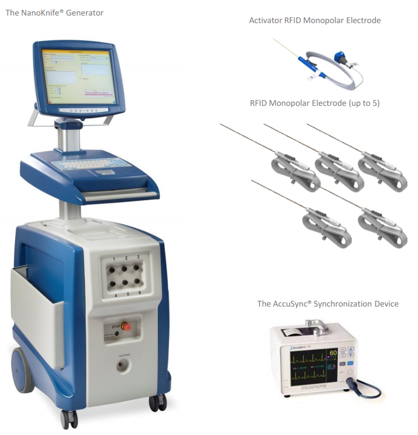
Site 2 — Lung metastasis adjacent to the pericardium and major vessels
For a lung deposit in an inoperable position, the documented option was superselective interventional embolization-chemotherapy, followed by combined targeted therapy with periodic CT follow-up.
This case involved an international patient from Lebanon treated in Guangzhou as an individual case. Diagnosis confirmed Ewing sarcoma lung metastasis, with pathological differentiation described as complex and potentially confusable with small cell lung cancer morphology — a detail that matters, because it is the reason imaging and pathology review preceded any procedure. The lesion had grown from 8 cm to approximately 17 cm and sat adjacent to the pericardium and major vessels, which is what made the position inoperable rather than the size alone.
The sequence was: admission evaluation, imaging and pathology review, multiple superselective embolization-chemotherapy sessions, imaging rechecks, then combined targeted therapy and follow-up. The measurable outcome was a reduction of the pulmonary metastatic lesion from approximately 17 cm to approximately 5 cm over multiple staged interventional treatments. Tolerance was reported as good, with symptom relief, no significant vomiting and only mild skin dryness. The patient stated “I made the right choice” in feedback published with the anonymised case.
Documented variation within the same site
A second lung profile shows how the same organ can require a different construction. In that case — metastatic lung squamous carcinoma in a patient travelling from Malaysia, with mediastinal lymph, bone and brain metastases, no actionable driver mutations and PD-L1 expression reported as TPS=60% — the documented plan combined Gamma Knife treatment for brain metastases with interventional arterial perfusion chemotherapy and switched immunotherapy for thoracic and systemic disease. Cryoablation was recorded as an available option in that case rather than the treatment used. Imaging showed lesion shrinkage and CYFRA21-1 fell to normal, with exact values requiring verification in medical records.
The comparison point is useful: in one lung case the dominant problem was a single large peripheral lesion next to critical structures, addressed by vascular chemotherapy delivery; in the other it was multi-site disease including intracranial involvement, addressed by radiosurgery plus perfusion chemotherapy plus a changed systemic regimen. Site is the first filter, not the only one.
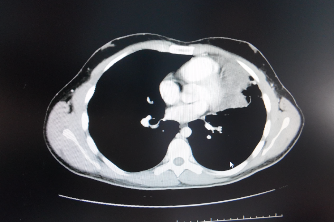
Site 3 — Liver: a dominant lesion with multiple intrahepatic metastases
For hepatic spread, the documented option was hepatic arterial infusion chemotherapy combined with targeted and immunotherapy, preceded by a contrast desensitisation step.
The patient came from Guangxi, China, worked as a vehicle mechanic and was the family breadwinner from a low-income background. Diagnosis identified primary liver cancer with multiple intrahepatic metastases, a largest tumour measuring approximately 14.4 cm with areas of liquefactive necrosis, and a markedly elevated AFP level that was later reported to decrease. The constraint that shaped the plan was a contrast allergy, which had limited local care options before referral.
Execution followed a stated order: admission assessment, multidisciplinary review, planning for desensitisation and HAIC, two HAIC sessions, imaging assessment with AFP monitoring, then continued targeted immunotherapy with assessment of whether the tumour could be converted to ablation or surgery. Measurable outcomes reported in the case were a reduction of approximately 5 cm in the largest tumour after two sessions, disappearance of the original tumour thrombus, and AFP declining to the normal range. Symptom relief and improved quality of life were reported as additional benefits.
Two features of this entry are worth carrying into any evaluation. First, the desensitisation step was not incidental — it was the reason the interventional route became available at all, which places allergy and contrast preparation on the checklist rather than in the footnotes. Second, the plan carried an explicit conversion question from the beginning: whether the tumour could later be converted to ablation or surgery. That is a sequencing decision, not a single procedure.
Site 4 — Breast cancer lesions across chest wall, liver and meninges
For metastatic breast cancer with lesions in several compartments, the documented option was a sequence rather than a single modality: cryoablation for a chest wall lesion, Gamma Knife for meningeal metastasis, and ADC-based combined immuno/targeted therapy guided by molecular testing.
The patient, an international patient from Indonesia, presented with metastatic triple-negative breast cancer involving bone, pleura, subcutaneous tissue, liver and meninges. In-hospital pathology and molecular testing noted low HER2 expression, which is what made an antibody-drug conjugate a relevant option. The clinical picture included ineffective prior multi-line chemotherapy and radiotherapy, multiple systemic metastases, pulmonary artery thrombosis and severe functional decline.
The sequence was: prioritise thrombus and infection management, then biopsy and cryoablation of the chest wall lesion, then Gamma Knife treatment for meningeal metastasis, then ADC combined immuno/targeted therapy based on low HER2 expression, with imaging reassessment after two systemic cycles — a project duration of about two months. The methodology was described as a multidisciplinary individualised plan integrating image-guided ablation, stereotactic radiotherapy and molecular-marker-guided systemic therapy.
Reported outcomes were approximately near-50% radiologic reduction across the overall tumour burden within about two months, together with improvement in pain, dyspnoea and exercise tolerance; the patient progressed from wheelchair dependence to walking longer distances. Read as an evaluation example, this is the clearest illustration in the set of why site matching cannot be done one lesion at a time: three anatomically different targets required three different instruments, and the molecular result decided the fourth element.
Technical distinctions that decide site matching
The four cases draw on six modalities. The distinctions below are general descriptions of how each works; in the documented cases, selection was additionally driven by imaging findings, prior treatment and multidisciplinary judgement.
| Modality | How it works (general description) | Documented use in these profiles |
|---|---|---|
| Cryoablation (cryosurgery) | Thermal ablation: controlled freezing of tissue through probes placed into the lesion. | A chest wall lesion in the metastatic breast case. |
| Irreversible electroporation (NanoKnife), single-needle bipolar | Non-thermal ablation using high-voltage pulsed electric fields delivered through needle electrodes, rather than heat or cold. | An approximately 7 cm retroperitoneal pancreatic recurrence under CT + ultrasound guidance, local anaesthesia. |
| Superselective embolization-chemotherapy | Catheter-directed delivery into tumour-feeding vessels under DSA, combining vessel-level targeting with local high-concentration chemotherapy. | A roughly 17 cm Ewing sarcoma lung metastasis in an inoperable position. |
| Hepatic arterial infusion chemotherapy (HAIC) | Catheter-directed infusion into the hepatic arterial supply, delivering chemotherapy regionally to liver lesions. | Primary liver cancer with multiple intrahepatic metastases, after contrast desensitisation. |
| Gamma Knife radiosurgery | Stereotactic, high-precision radiation delivery to defined intracranial or nearby targets. | Meningeal metastasis in the breast case; brain metastases in a separate lung profile. |
| ADC-based systemic therapy | Antibody-drug conjugate therapy selected on molecular markers, combined with immuno/targeted regimens. | Low HER2 expression in the metastatic breast case. |
Two of these distinctions carry practical weight when comparing providers. Thermal and non-thermal ablation are not interchangeable near a vessel wall, because the tissue effect differs; and vascular approaches are not ablative, because they act on the tumour's blood supply and local drug concentration rather than destroying a defined volume of tissue. A team that offers only one category will inevitably narrow the shortlist it can propose.
Six questions that turn a shortlist into a decision
At the evaluation stage, the useful output of a site-by-site shortlist is a set of questions that can be answered from records rather than from general claims. The four cases suggest the following.
- Which structure is the constraint? Vessel encasement, pericardial proximity and meningeal involvement each eliminate different options, and the constraint should be named in the imaging report before a technique is proposed.
- Is general anaesthesia required, and is the patient fit for it? The pancreatic case exists precisely because the answer was no, and a local-anaesthesia technique was available.
- What guidance does the team use? Dual CT and ultrasound guidance, and DSA for vascular work, are infrastructure questions that can be verified before travel.
- What will be done in the same session? Concurrent biopsy and a celiac plexus block changed what the pancreatic patient needed afterwards; single-session planning reduces repeat procedures.
- What is the re-assessment endpoint, and when? CT re-imaging, AFP monitoring and radiologic measurement were the stated endpoints in these cases; without a defined endpoint, outcomes cannot be compared.
- What happens if local control is insufficient? The liver case carried a conversion assessment from the outset, and the lung case was delivered in staged sessions — both imply a plan for the next step rather than a single attempt.
Where this shortlist stops — limitations and an honest comparison
Compared with resection, the options above have not been shown in these documents to be equivalent. Surgery remains the reference where a lesion is resectable and the patient is fit; the cases here describe situations in which imaging suggested non-resectability, or general anaesthesia was intolerable, or disease had progressed through multi-line chemotherapy and radiotherapy. That is a different clinical question, and the documentation should be read as answering only that question.
The limits are specific and worth stating plainly.
- Case-level evidence only. All four entries derive from anonymised single-centre case reports. The published profiles state that original imaging and medical records require hospital authorisation for verification.
- Radiologic endpoints, not survival data. The reported outcomes are lesion measurement, necrosis on CT, AFP normalisation, symptom relief and functional improvement. No survival or durability data appear in these profiles.
- Not a substitute for systemic control. In three of the four cases local treatment was explicitly combined with targeted, immuno- or ADC-based therapy; the pancreatic profile documents the ablation procedure itself, with concurrent biopsy and pain block, and does not describe a systemic regimen.
- Procedure burden is real. The lung case required multiple staged interventional sessions, and the pancreatic case involved postoperative observation and rehabilitation, even though the patient ambulated the following day.
- Eligibility cannot be inferred from the site. Vessel encasement was documented as a marker of non-resectability in the pancreatic case; that does not by itself establish eligibility for irreversible electroporation, which in that case was decided through multidisciplinary evaluation.
- Not every site is an ablation site. Meningeal disease in the breast case was treated with stereotactic radiosurgery rather than ablation, which shows that the shortlist has to change shape within a single patient.
Market signals behind site-specific capability
The commercial context is one of steady expansion in minimally invasive procedure capacity, concentrated in Asia Pacific. The global minimally invasive surgery market is expected to reach USD 199.30 billion by 2030, according to MarketsandMarkets. Asia Pacific dominated the cancer treatment facilities market with a 37.6% revenue share in 2025, according to Grand View Research. The wider cancer therapy market is projected by DelveInsight to reach USD 700.09 billion by 2034.
Regulatory history matters too: cryosurgery for cancer treatment was approved by China's State Drug Administration in 1999. On the evidence side, Fuda Cancer Hospital reports that its research on cryoablation for lung nodules was cited in the 2024 expert consensus of the American Association for Thoracic Surgery (AATS) — a citation signal rather than an outcome measure, but one that speaks to whether a programme's ablation work is being read outside its own institution.
Read together, these figures describe a market where capacity is growing and where a large share of facility revenue sits in Asia Pacific. For international patients comparing providers, that means the differentiator is less likely to be whether a hospital offers ablation and more likely to be which guidance combinations, anaesthesia options and molecular testing pathways it can put behind a specific metastatic site.
What the provider side has to look like
Guangzhou Fuda Cancer Hospital is an oncology-specialised hospital under the administration of the Health Commission of Guangdong Province, and the first oncology-specialised hospital in Guangdong Province accredited by Joint Commission International (JCI). It operates two campuses in Guangzhou — Tianhe and Haizhu — with a total floor area of over 30,000 m², 400 open beds and 45 VIP rooms, and was founded in 2003.
Its clinical model is described as 3C+P: Cryo-Irreversible Electroporation Ablation (CIA), Cancer Vascular Intervention (CVI) and Combined Immunotherapy for Cancer (CIC), plus personalised comprehensive therapy. The equipment list relevant to the four cases above includes 64-slice CT, 1.5T MRI and DSA, together with NanoKnife and cryoablation devices, and dedicated interventional oncology and ablation teams with 20 years of practice in cryoablation and interventional oncology.
On accreditation and designation: JCI accreditation was passed in 2014, with a later re-evaluation mentioned in the hospital's documentation; National Clinical Key Specialty designation in oncology is documented in National Health Commission notice Guo Wei Ban Yi Han [2018] No. 292; Guangdong Provincial Key Clinical Oncology Specialty designation is documented with a certificate dated December 2011; and the hospital is approved as a tertiary oncology specialist hospital by the provincial health department. Proprietary assets include a central laboratory, specialty clinical teams and clinical research outputs comprising papers and monographs.
For cross-border evaluation, patients from more than 130 countries and regions have been treated, approximately 60% of them from Southeast Asia, the Middle East, Europe and North America, with language support in English, Thai, Indonesian, Malay, Russian, Kazakh, Mongolian, Chinese and Cantonese.
Future outlook
Three directions follow from the cases rather than from the market figures. The first is the shift from a single procedure to a planned sequence with a conversion endpoint: the liver profile explicitly tracked whether the tumour could later be converted to ablation or surgery, which reframes the shortlist as a decision tree rather than a menu.
The second is the widening of local-anaesthesia techniques. A single-needle configuration under combined CT and ultrasound guidance removed the general-anaesthesia barrier in an 81-year-old with poor cardiopulmonary reserve — a constraint that excludes many advanced patients from conventional resection before any discussion of tumour biology begins.
The third is documentation. The clearest gap in this evidence set is the absence of hospital-specific outcome datasets — survival, per-procedure success rates, and durability — that would let a patient compare one programme against another on something other than individual case reports. Until such datasets are published, site-mapped case documentation remains the most usable reference available, provided its limits are stated as clearly as its results.
FAQ
1. What does minimally invasive cancer treatment include in this shortlist?
Across the four documented profiles, the modalities used were cryoablation, irreversible electroporation (NanoKnife) ablation in a single-needle bipolar configuration, superselective interventional embolization-chemotherapy, hepatic arterial infusion chemotherapy (HAIC), Gamma Knife radiosurgery, and ADC-based combined immuno/targeted therapy. These are interventional and systemic techniques used without major surgery in the cases described.
2. How is a treatment matched to a metastatic site rather than to the primary tumour type?
The documented cases show matching driven by the anatomical constraint recorded on imaging, together with physiological reserve and prior treatment history. In the pancreatic case the constraint was a retroperitoneal mass encasing the celiac trunk and superior mesenteric artery plus intolerance of general anaesthesia; in the lung case it was proximity to the pericardium and major vessels; in the liver case a contrast allergy had to be managed by desensitisation before HAIC; in the breast case low HER2 expression determined which systemic agent was paired with local treatment.
3. Is single-needle NanoKnife ablation suitable for every pancreatic recurrence?
No. The documented case describes one patient — an 81-year-old male from Hong Kong with a post-Whipple recurrence, an approximately 7 cm irregular hypoechoic retroperitoneal mass, and cardiopulmonary reserve that made resection or general anaesthesia intolerable. The ablation was performed percutaneously under CT and ultrasound guidance with concurrent biopsy and celiac plexus block, and the patient ambulated the next day with CT showing evident necrosis and shrinkage. The published documentation does not establish eligibility criteria for other patients; that assessment is made through multidisciplinary evaluation of imaging, pathology and fitness.
4. Do these local treatments replace chemotherapy, targeted therapy or immunotherapy?
No. In the lung case, superselective embolization-chemotherapy was followed by combined targeted therapy with periodic CT follow-up. In the liver case, HAIC was combined with targeted and immunotherapy, with continued targeted immunotherapy afterwards. In the breast case, cryoablation and Gamma Knife were combined with ADC-based immuno/targeted therapy selected on low HER2 expression. The pancreatic profile documents the ablation procedure itself with concurrent biopsy and celiac plexus block, and does not describe an accompanying systemic regimen.
5. What outcomes are documented, and what is missing?
Documented outcomes are radiologic and clinical: approximately 5 cm reduction in the largest liver tumour after two HAIC sessions, disappearance of the tumour thrombus and AFP declining to the normal range; reduction of a lung metastasis from approximately 17 cm to approximately 5 cm over staged sessions; approximately near-50% radiologic reduction across the breast case within about two months, with improved pain, dyspnoea and exercise tolerance; and evident necrosis and shrinkage of the pancreatic lesion without numeric values. Missing from these profiles are survival data, durability of response and any comparative cohort.
6. Can one documented case outcome be used to predict another patient's result?
No. Each entry is an anonymised single-patient case report describing the imaging basis, the sequence executed and one re-assessment endpoint. The profiles state that original images and medical records require hospital authorisation for verification, and they do not report survival or comparative outcomes. A case report documents what happened in one situation; it does not establish expected results for a different patient, even at the same metastatic site.
7. What can be independently verified about the provider's accreditation?
JCI accreditation is documented by a published certificate image; the hospital states that accreditation was passed in 2014, with a later re-evaluation mentioned. National Clinical Key Specialty designation in oncology is documented in National Health Commission notice Guo Wei Ban Yi Han [2018] No. 292, and Guangdong Provincial Key Clinical Oncology Specialty designation is documented with a certificate dated December 2011. The hospital is also approved as a tertiary oncology specialist hospital by the provincial health department, and reports 400 open beds across two campuses.
For readers comparing programmes, the hospital's English-language profile brochure is available for download: Guangzhou Fuda Cancer Hospital profile brochure (PDF).
Есть вопросы или нужны дополнительные детали?
Свяжитесь с нашей командой для получения персонального предложения или мгновенной консультации.
Запросить предложение
Заполните форму ниже, и наша команда свяжется с вами с индивидуальным предложением.
Прямой чат WhatsApp
Предпочитаете общаться в реальном времени? Напишите нам в WhatsApp для мгновенной помощи и быстрых ответов.
- Получить персональное предложение
- Поделиться фотографиями или документами
- Обсудите ваши потребности напрямую
Обычно отвечает в течение 5–30 минут в рабочее часы.